News
Throwing Light on STEM Innovation
September 4, 2018
Pictured: Scan of a domestic chicken at 0.037 mm resolution, smaller than the width of a strand of hair.
NYIT researchers have been awarded a grant for $426,621 from the National Science Foundation (NSF) for a micro-computed tomography machine—a high-resolution device used for microscopic imaging.
Also known as a micro-CT scanner, the machine uses x-ray imaging technology (similar to CAT scans used in hospitals) to make interior structures of objects and specimen visible at a microscopic level. Researchers at NYIT will use the device to explore the internal morphology of fossils, measure bone density, map the inner structure of the brain, and closely examine intricate vessels and other soft tissue structures—tasks that without the machine require dissection and often cause irreversible damage to specimen.
The machine will be housed in NYIT College of Osteopathic Medicine’s (NYITCOM) Data Visualization Lab on the Long Island (Old Westbury, N.Y.) campus, and will be available free of charge to both faculty and student researchers from NYIT as well as outside institutions.
The micro-CT scanner’s accessibility is expected spark collaboration between NYIT faculty and researchers from neighboring institutions, supporting NYIT ’s innovative culture and contributions to the Greater New York research community. Lead investigator Simone Hoffmann, Ph.D., assistant professor of anatomy, emphasized the impact of the grant, which will enable NYIT to become one of the region’s few institutions to offer micro-CT scanning capabilities without a fee.
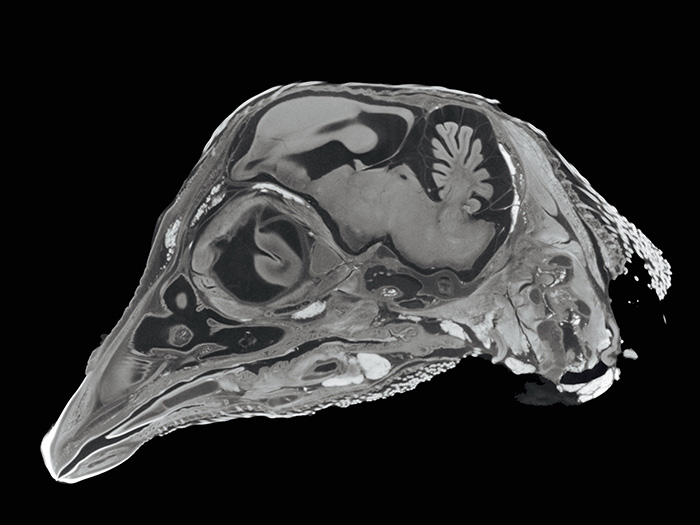
Image of a chicken highlights a technique called diffusible iodine contrast-enhanced (dice) CT imaging, which allows researchers to visualize and reconstruct soft tissue in 3-D and high-resolution. This process allows scientists to differentiate between soft-tissue structures in x-ray images without damaging the specimen.
“As a researcher, it’s very difficult to complete your study when each scan comes with a price tag,” said Hoffmann. “Increased accessibility to this technology will help establish NYIT as a central research facility on Long Island and nurture innovative research in biology, paleontology, nanotechnology, engineering, and life sciences.”
Researchers from NYIT College of Engineering and Computing Sciences and NYIT College of Arts and Sciences, including Assistant Professor of Electrical and Computer Engineering Azhar Ilyas, Ph.D., are also eager to begin using the technology. Ilyas, who was a co-investigator for the grant, will use the new micro-CT scanner to study bone healing.
“While current data partially explains the complex biological pathways through which bone is regenerated, much remains to be understood about the host tissue interactions and fracture repair process. The acquisition of this new imaging technology will allow us to answer many of those questions,” said Ilyas.
Students will also have new opportunities for experiential learning. Engineering students, led by faculty, will integrate the CT-based technology in the New York State Industries for the Disabled Cultivating Resources for Employment with Assistive Technology (CREATE) program, which develops technologies to assist people with disabilities in their work environments. Similarly, College of Arts and Sciences faculty will lead students in examining the 3-D data retrieved during scanning.
Other grant co-investigators included Olga Savinova, Ph.D., assistant professor of biomedical sciences, Julia Molnar, Ph.D., assistant professor of anatomy, and Claude Gagna, Ph.D., associate professor of life sciences.
The scanner is expected to be available in early 2019.