Imaging Core Facility
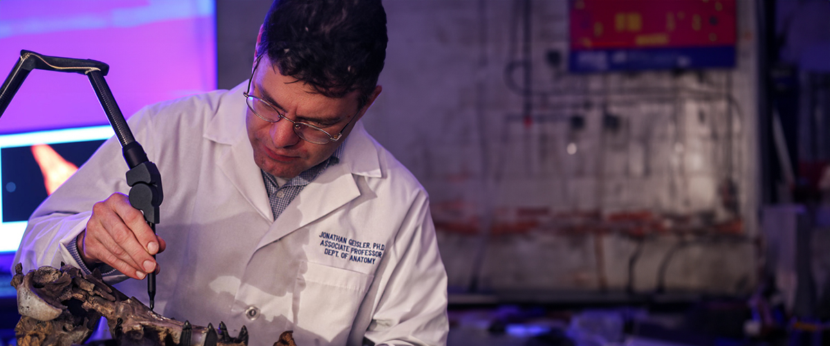
The imaging facility offers an array of digital imaging microscopy systems to assist our medical school researchers in their analysis of fossils, tissue, and cells at the macroscopic, cellular, and sub-cellular levels. Image processing software associated with the confocal microscopes and the Sensofar S Neox optical profiler system enables the 3-dimensional and 4-dimensional reconstruction of biological samples and analysis of the sample images. Student access is limited to students participating in related faculty-led research projects.
Equipment contained within the Imaging Core Facility includes:
- Sensofar S Neox optical profiler with SensoSCAN and SensoMAP software to analyze 3D and surface properties of samples.
- Multiple upright and inverted fluorescence microscopes with imaging software.
Location
Kenneth J. Riland Academic Health Care Building, NYITCOM-Long Island